Professor Charmaine Childs
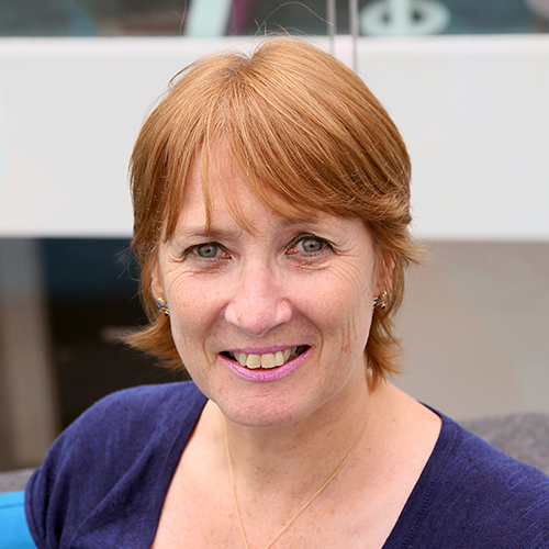
Charmaine Childs is Professor of Clinical Science in the Health Research Institute, Sheffield Hallam University. Charmaine leads the Applied Imaging in Health and Disease research programme. Her strategic research objective is research translation into practice via development of new technology at the bedside/clinic.
Charmaine has held senior research and academic positions in the United Kingdom (Medical Research Council, UK from 1991-2000) and the School of Medicine University of Manchester, UK from 2000-2009 followed by work overseas at the Yong Loo Lin School of Medicine, National University of Singapore (2009-2013).
Her work with a diverse group of scientists, engineers, computer scientists, architects and clinicians has contributed to a rich experience with international cross-disciplinary collaborations.
Her work in surgical site infection imaging is recognised internationally, winning four international awards.
Presentation at The Society of Tissue Viability 2023 Conference
New approaches to surgical site infection and cutaneous perfusion imaging using infrared thermography
Objectives
After attending this session, persons will be able to:
- Understand the basic principles of infrared radiation applied to thermography
- Methods and surrogates of perfusion assessment
- Perfusion assessment in the peri-opeative period
- New research in wound management using dynamic infrared video thermography
Abstract
Living tissue is subjected to many noxious insults both accidental and deliberate. Being able to make an accurate and reliable assessment of a wound is seldom straightforward. In this lecture skin and wound imaging will be discussed with respect to non-inasive imaing modalities. The main focus will be on surgical incision, a procedure which disrupts delicate skin and subcutaneous structures. Whilst the procedures are beneficial for the health and wellbeing of the patient, there is little doubt that exposure of internal structures increases the risk of contamination and infection by pathogens.
Typically, infection is not obvious until changes at the incision site are visible by eye but what if more information could be provided by examining the wound in a different light spectrum; long wave infrared?
The aim of this lecture is to provide a background to the science of medical thermography, exploring new methods for skin perfusion imaging and the opportunities for objective wound assessment that can be delivered at the bedside.
Advances in surgical wound management and reducing Surgical Site Infection (SSI) advanced study day
Progress in wound imaging: what place for infrared thermography in assessment of surgical wounds?
Abstract
Living tissue is subjected to many noxious insults both accidental and deliberate. Surgical incision disrupts delicate skin and subcutaneous structures and whilst the procedures are beneficial for the health and wellbeing of the patient, there is little doubt that exposure of internal structures increases the risk of contamination and infection by pathogens.
Typically, infection is not obvious until changes at the incision site are obvious to the human eye but what if more information could be provided by examining the wound in a different light spectrum; long wave infrared?
The aim of this lecture is to explore opportunities for objective wound assessment using imaging to detect subtle early-stage signatures of tissue changes using a human surgical model; caesarean section.

